Microscopy in Bone
Bone is a very dense and opaque tissue that is difficult to see into. Other tissue types like skin allow more light to penetrate, making them more amenable to visualizing with normal methods such as confocal microscopy. Think of two glasses filled with liquid: skin is like lemonade, easy to see through, while bone is like milk or even mud, a task much more difficult.
Different types of microscopy have been used to interrogate bone in the past, but most methods involve taking the bone outside of its normal, living environment, and sometimes even dissolving the mineral of the bone so the cells could be seen. This greatly disrupts the microenvironment of cells, which is known to have a critical impact on their function. Cells don’t perform the same way when you change the home that they live in. So it has been necessary to develop novel methods on imaging cells inside of living bone.
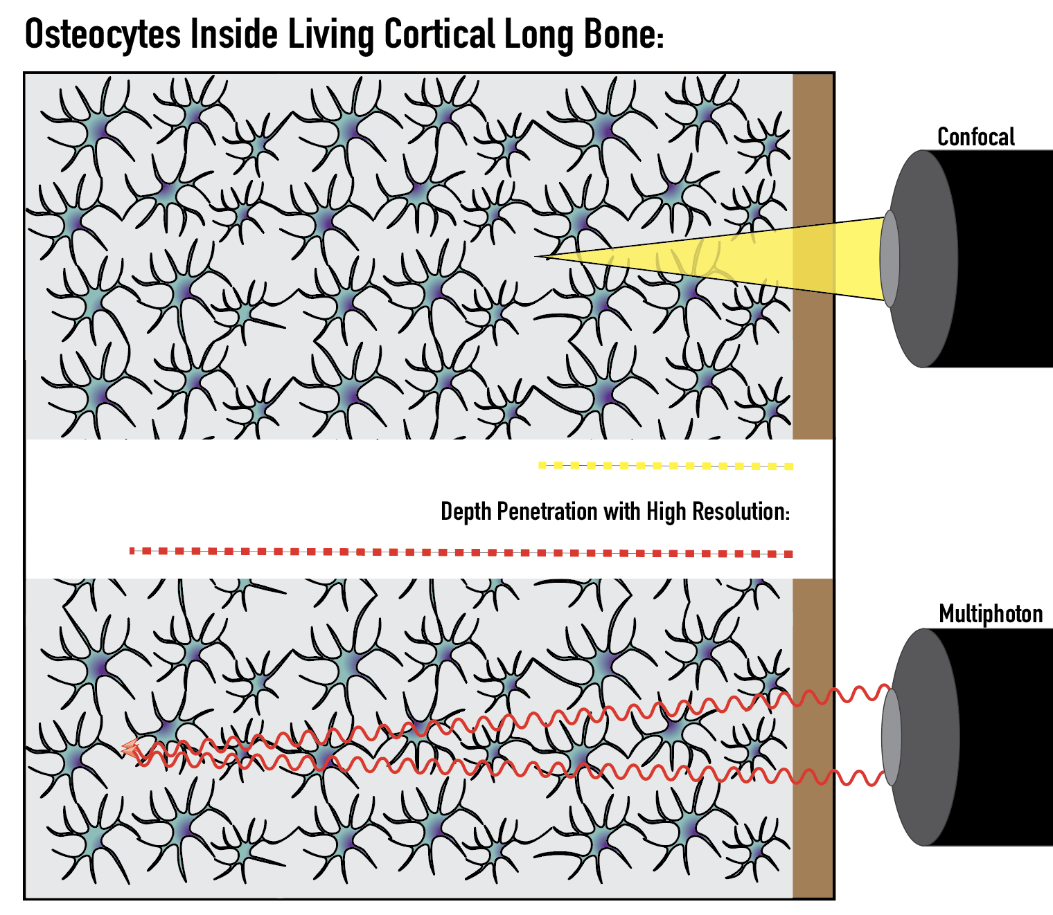
Recent advancements in a powerful type of microscopy, multiphoton microscopy, have allowed cells inside living bone to be visualized deeper with high resolution. This means that fine structures of osteocytes and their neuron-like dendrites, can be seen at depth into a bone!
Instead of illuminating an entire cone of the tissue (picture a flashlight beam), multiphoton microscopy sends two beams of individual photons into the tissue at rapid succession (picture two small lasers meeting at a pinpoint). This physics discovery makes opaque tissue like bone accessible for researchers to see into without making large modifications to the tissue environment.
With these advancements, new questions can be asked about the structure and function of osteocytes inside of living bone, with intact fluid flow and microarchitecture, which is a large part of what we do in the Lewis Lab!
